


The traditional concept of how the human brain governs voluntary movement may not be accurate.
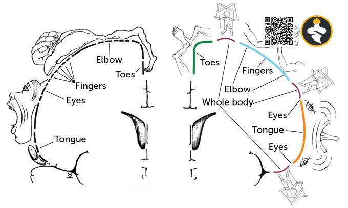
The motor homunculus map of the primary motor cortex demonstrates how this brain area is split into portions assigned to each body part that may be manipulated freely. It aligns your toes with your ankle and your neck with your thumb. The amount of space each part occupies in the cortex is related to how much control one has over that part. Each finger, for example, occupies more area than a whole thigh.
According to a new map, three additional zones govern integrative, whole-body activities, in addition to sections dedicated to individual body parts.
Researchers report in Nature on April 19 that representations of where certain body parts lie on this map are arranged differently than previously imagined.
This had been suggested by monkey research. “There’s a whole cohort of people who have known for 50 years that the homunculus isn’t quite right,” says Evan Gordon, a neurologist at Washington University in St. Louis. However, since neurosurgeon Wilder Penfield’s pioneering brain-mapping work in the 1930s, the homunculus has ruled supreme in neuroscience.
Gordon and his colleagues are interested in coordinated activity and communication between brain areas.
They discovered that some parts of the primary motor cortex were related to previously unknown areas involved in movement control and pain perception. They dismissed it as a product of faulty data since it did not match the homunculus map. “But we kept seeing it, and it was bugging us,” Gordon adds.
As a result, the researchers collected functional MRI data on participants while they performed various tasks.
Two participants performed basic actions such as moving their brows or toes, as well as sophisticated activities such as twisting their wrists and moving their feet from side to side at the same time.
The fMRI data indicated which portions of the brain responded concurrently with each task, allowing the researchers to determine which regions were functionally related to one another. Seven more subjects were videotaped while not performing any activity in order to examine how brain regions communicate when at rest.
Gordon claims that testing only a few subjects for several hours each provides unique insights into brain connections. “When we collect this much data from individuals, we constantly start seeing things that people have never really noticed before.”
While the brain-body part connections follow a pattern similar to that established by Penfield, the main motor cortex is divided into three different parts. Each one depicts a distinct part of the body:
The lower body, chest, limbs, and head are all visible.
The outermost body component of that area is mapped to the center of that segment inside each of these parts. For example, the toes are in the midst of the major motor cortex region assigned to the lower body, with additional leg components radiating out in each direction from it. As a result, the portion is structured as follows: hip, knee, ankle, toes, ankle, knee, hip.
The scientists also discovered three unexplained areas that were not associated with any bodily organ. They are known as inter effector regions because they link to an external network that is engaged in action control and pain sensing. These sections alternate with those dedicated to specific bodily components.
The team estimates that inter-effector areas may combine action objectives and multiple-body-part motions, whereas the gaps in between are employed for precise movements of isolated body parts.
The scientists confirmed that this arrangement was constant across a wide range of participants by using prior data from three big fMRI investigations, which included data from about 50,000 people. Existing datasets from macaque monkeys, children, and clinical populations revealed similar findings.
“I think it was just easy to miss anything that seemed anomalous — must be noise,” says Michael Graziano, a Princeton University neurologist who was not involved in the study.
But now that these massive datasets are available, “you have these tremendous numbers of individuals, and the pattern is crystal evident, and you can’t ignore it…. This is the greatest example I’ve seen in a long time of looking at folks and trying to figure out what the organization is at a detailed level.”
Gordon’s team will now investigate whether these inter effector areas are involved in particular types of pain. More broadly, the team hopes that their results will spur further investigation into what certain parts of the brain do. Gordon believes that with new techniques and tools, there is still plenty to discover. “Brain mapping isn’t dead.”
Reference: Nora Bradford@sciencenews.org